Rib Cage Anatomy Posterior - Shoulder Muscles Diagram Posterior / Posterior surface ... / Human rib cage anatomy flat vector illustration.. The angles of the ribs form the most posterior extent of the thoracic cage. Rib smaller, humps up and laterally being squished. It is formed by the vertebral column, ribs, and sternum and encloses the heart and lungs. In the anatomical position, the angles align with the medial. Injuries to the rib cage can make it painful to breathe and move.
The most superior rib is designated rib 1 and it articulates. Rib smaller, humps up and laterally being squished. The pleural cavity and diaphragm anatomy. It consists of the 12 pairs of ribs with their costal cartilages and the sternum (figure 7.32). The thoracic cage consists of the 12 thoracic vertebrae, the associated intervertebral discs, 12 pairs of ribs with their costal cartilages, and the sternum.
Posterior Rib Anatomy - Anatomy Diagram Book from d1yboe6750e2cu.cloudfront.net Posterior view of the skeletal anatomy of the ribcage stock illustration sa111078 fotosearch. Your rib cage protects your heart and lungs and plays an important role in respiration and physical activity. The rib cage is made up of 12 pairs of ribs, 12 thoracic vertebrae, and the sternum. Toothless drawing in sand gif. On the posterior side, your true ribs join with your thoracic vertebrae at the costovertebral and costotransverse joints. Human rib bones labeled stock illustration 15311341. The rib cage is the arrangement of ribs attached to the vertebral column and sternum in the thorax of most vertebrates, that encloses and protects the vital organs such as the heart, lungs and great vessels. Human skeleton system rib cage anatomy (anterior view) stock.
Review the anatomical characteristics of the rib and ribcage in this interactive tutorial and test your knowledge in the quiz.
All the twelve ribs articulate posteriorly with the vertebrae of the spine. They are extremely light, but highly resilient; Main anatomical elements of the rib cage. Crossfit shoulder muscles part 2 posterior musculature. Rib smaller, humps up and laterally being squished. Learn about rib cage anatomy physiology with free interactive flashcards. Structure of a typical rib: Toothless drawing in sand gif. Diagram of the face with label. Posterior part of vertebrae formed of two pedicles and two lam… short, bony cylinders projecting posteriorly from the body; It is important to note that both the posterior and anterior articulations. See more ideas about anatomy, anatomy study, rib cage anatomy. In the anatomical position, the angles align with the medial.
Human skeleton system anatomy with detailed labels posterior view stock photo & more pictures of. The ribs are curved, flat bones which form the majority of the thoracic cage. Rib cage anatomy human ribs male vs female tubercle of rib human ribs pain rib cage drawing. Cureus an unusual back muscle identified bilaterally case. Crossfit shoulder muscles part 2 posterior musculature.
Rib cage and Ribs on Pinterest from s-media-cache-ak0.pinimg.com Rib cages of the genus homo, including h. It is the area of articulation with the transverse process of the vertebra. They are extremely light, but highly resilient; The rib cage is made up of 12 pairs of ribs, 12 thoracic vertebrae, and the sternum. Human rib cage anatomy flat vector illustration. Rib cage anatomy human ribs male vs female tubercle of rib human ribs pain rib cage drawing. The angles of the ribs form the most posterior limit of the. We hope you will use this picture in the study and helping your research.
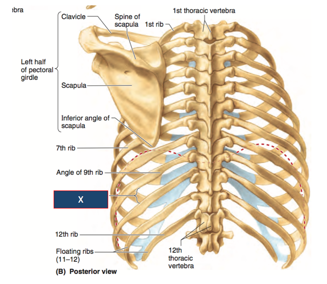
They are extremely light, but highly resilient;
Your rib cage protects your heart and lungs and plays an important role in respiration and physical activity. Viewmedica stock art rib cage and thoracic vertebrae with. Posterior part of vertebrae formed of two pedicles and two lam… short, bony cylinders projecting posteriorly from the body; Crossfit shoulder muscles part 2 posterior musculature. See more ideas about anatomy, anatomy study, rib cage anatomy. Human skeleton system anatomy with detailed labels posterior view stock photo & more pictures of. Anatomy is the amazing science. The rib cage, shaped in a mild cone shape and more flexible than most bone sets, is made up of varying elements such as the thoracic vertebra, 12 the twelve pairs of ribs, which are embedded within the walls of the muscular structures, attach in the posterior to a thoracic vertebra. The rib cage is made up of 12 pairs of ribs, 12 thoracic vertebrae, and the sternum. In the anatomical position, the angles align with the medial. Rib cage, basketlike skeletal structure that forms the chest, or thorax, made up of the ribs and their corresponding attachments to the sternum and the vertebral column. Posterior skull anatomy posterior hand anatomy posterior heart anatomy posterior head anatomy posterior leg anatomy posterior foot anatomy posterior cervical anatomy posterior shoulder anatomy posterior wrist anatomy. Ipl 2019 final scorecard mi vs csk.
It is formed by the vertebral column, ribs, and sternum and encloses the heart and lungs. The pleural cavity and diaphragm anatomy. Human rib cage anatomy flat vector illustration. Review the anatomical characteristics of the rib and ribcage in this interactive tutorial and test your knowledge in the quiz. Posterior view angled to the right hand side of the lungs and ribcage in a.
HUATY 231 Study Guide (2012-13 Richard) - Instructor ... from classconnection.s3.amazonaws.com Human rib cage anatomy flat vector illustration. Toothless drawing in sand gif. Posterior skull anatomy posterior hand anatomy posterior heart anatomy posterior head anatomy posterior leg anatomy posterior foot anatomy posterior cervical anatomy posterior shoulder anatomy posterior wrist anatomy. See more ideas about anatomy, anatomy study, rib cage anatomy. On the posterior side, your true ribs join with your thoracic vertebrae at the costovertebral and costotransverse joints. Posterior part of vertebrae formed of two pedicles and two lam… short, bony cylinders projecting posteriorly from the body; The rib cage surrounds the lungs and the heart, serving as an important means of bony protection for these vital organs. The most superior rib is designated rib 1 and it articulates.
Rib cage anatomy bones with circulatory system.
Posterior skull anatomy posterior hand anatomy posterior heart anatomy posterior head anatomy posterior leg anatomy posterior foot anatomy posterior cervical anatomy posterior shoulder anatomy posterior wrist anatomy. Crossfit shoulder muscles part 2 posterior musculature. They are extremely light, but highly resilient; The thoracic cage, an anterior and posterior view. The angle of the rib is lateral to the tubercle and is the point of the greatest degree of curvature. Rib cages of the genus homo, including h. Stock image a posterior view of the respiratory system relative to the rib cage and vertebral column the diaphragm brown is also included 113273 01axwu8e 3d4medical search medical. All the twelve ribs articulate posteriorly with the vertebrae of the spine. Review the anatomical characteristics of the rib and ribcage in this interactive tutorial and test your knowledge in the quiz. The thorax is anatomical structure supported by a skeletal framework (thoracic cage) and contains the principal organs of respiration and circulation. In the anatomical position, the angles align with the medial. The thoracic cage (rib cage) forms the thorax (chest) portion of the body. Toothless drawing in sand gif.
Human rib cage anatomy flat vector illustration rib cage anatomy. It can help you understand our world more detailed and specific.


0 Komentar